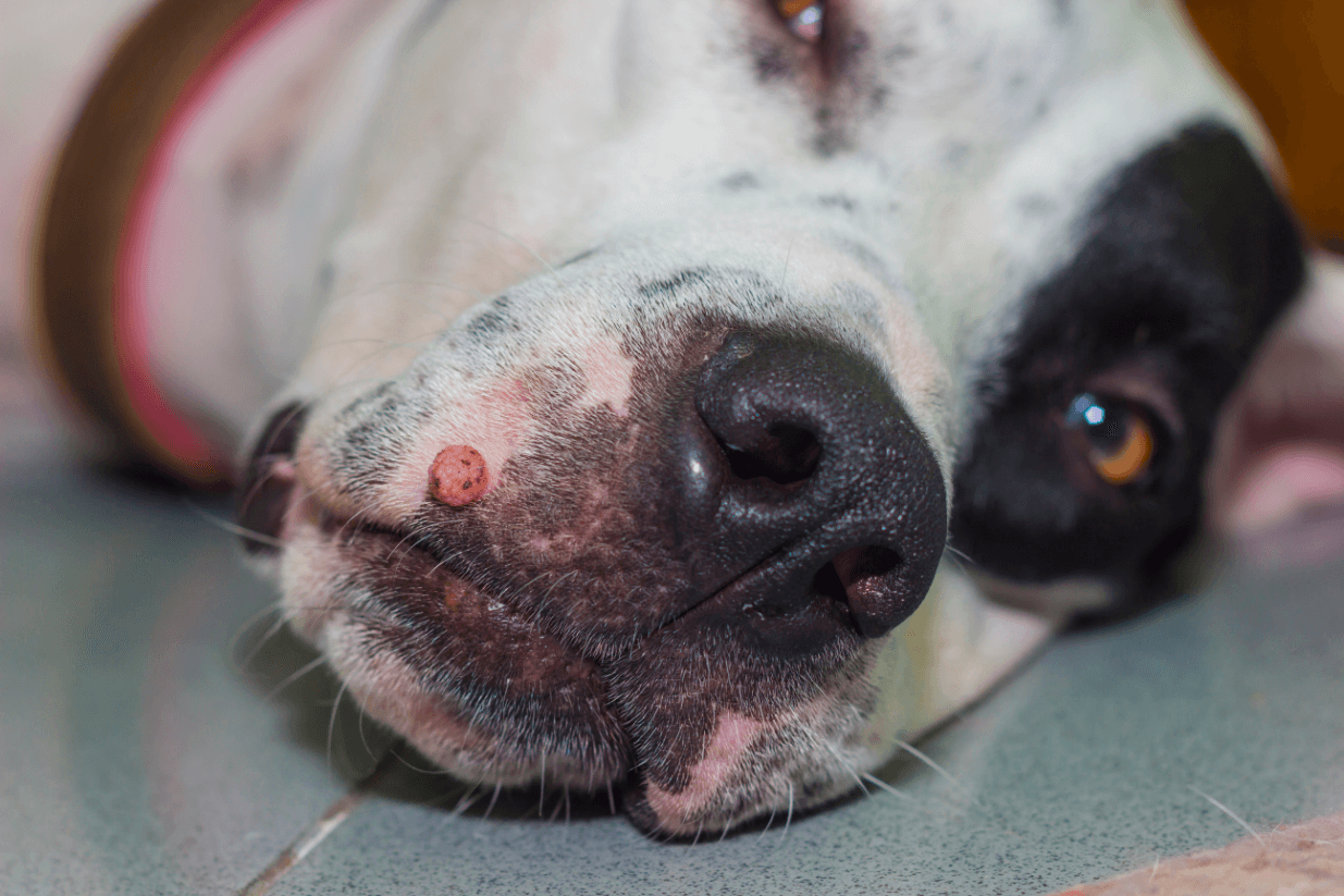
Dog Papilloma: Symptoms, Treatment, and Prevention Tips for Pet Owners
Discover essential insights on dog papillomas and learn how to identify, prevent, and treat these common warts. Ensure your pet’s health with expert advice today!
Have you ever noticed unusual growths in your dog’s mouth or on their skin? These may be papillomas, commonly known as dog warts. Canine papillomavirus (CPV) is a widespread issue affecting many of our furry friends, particularly young dogs.
We’ll discover the area of dog papillomas, from their causes to their symptoms and treatments. Understanding this condition is crucial for pet owners, as it can impact our dogs’ health and quality of life. While often harmless, these growths can sometimes lead to complications if left untreated. By exploring into the details of CPV, we’ll equip you with the knowledge to spot early signs and make informed decisions about your pet’s care.
What Is Dog Papilloma?
Dog papilloma, also known as canine papillomatosis, is a condition characterized by benign tumors or wart-like growths caused by the canine papillomavirus (CPV). These growths typically appear on a dog’s skin, mouth, or mucous membranes.
Causes and Transmission
Canine papillomavirus is the primary cause of dog papillomas. This double-stranded, non-enveloped DNA virus is highly host-exact, meaning it only affects dogs. Transmission occurs through direct contact with infected dogs or contaminated objects in the environment.
The virus requires microabrasions in the skin to access the basal layer and establish an infection. While visible lesions are the most common sign of infectiousness, it’s unclear if dogs without visible growths can transmit the virus.
Papillomas typically develop after a 4-week incubation period. But, subclinical infections may also occur, where dogs carry the virus without showing symptoms. Young dogs are particularly susceptible to oral papillomatosis, while cutaneous papillomas can affect dogs of any age.
Key points about CPV transmission:
- Direct contact with infected dogs
- Contact with contaminated objects
- Requires skin microabrasions for infection
- 4-week incubation period
- Possible subclinical infections
Understanding the causes and transmission of dog papillomas helps pet owners take preventive measures and recognize early signs of infection in their canine companions.
Types of Canine Papillomas
Canine papillomas manifest in various forms depending on their location and the exact papillomavirus strain causing the infection. The two primary types of canine papillomas are oral papillomas and cutaneous papillomas, each with distinct characteristics and implications for a dog’s health.
Oral Papillomas
Oral papillomas are the most common type of canine papilloma, typically caused by Canine Papillomavirus 1 (CPV1). These wart-like growths appear in the mouth, often on the lips, tongue, and palate. Young dogs are particularly susceptible to oral papillomatosis, with lesions developing rapidly and sometimes in clusters.
Characteristics of oral papillomas include:
- Appearance: Exophytic masses, single or multiple
- Location: Oral cavity, occasionally extending to the larynx and esophagus
- Duration: Usually regress spontaneously within 4-8 weeks
- Complications: Potential dysphagia or respiratory obstruction if many
Histologically, oral papillomas show:
- Papillary exophytic proliferation of squamous epithelium
- Koilocytosis
- Intranuclear inclusion bodies
While often benign, oral papillomas can interfere with eating and drinking if they become many or large. In most cases, they resolve on their own without treatment.

Cutaneous Papillomas
Cutaneous papillomas affect a dog’s skin and can appear anywhere on the body. These growths are less common than oral papillomas but can affect dogs of any age. Cutaneous papillomas are associated with various papillomavirus strains and may present differently depending on the exact virus and location.
Types of cutaneous papillomas include:
- Pedal papillomas:
- Occur on the footpads
- Less frequently studied than oral papillomas
2. Inverted papillomas:
- Associated with CPV1
- Grow inward rather than outward
3. Viral pigmented plaques:
- Linked to papillomaviruses of the Chi genus
- May progress to squamous cell carcinomas in some cases
Cutaneous papillomas typically appear as small, raised growths on the skin. They may be solitary or occur in clusters, and their appearance can vary from smooth and flesh-colored to rough and pigmented. While many cutaneous papillomas resolve on their own, some may persist or require treatment, especially if they cause discomfort or interfere with the dog’s normal activities.
In rare cases, certain types of cutaneous papillomas, such as viral pigmented plaques, have the potential to progress to malignant tumors. This underscores the importance of monitoring any persistent skin growths and consulting with a veterinarian for proper diagnosis and management.
Symptoms and Identification
Dog papillomas, also known as canine oral papillomas or dog warts, are benign growths caused by the canine papillomavirus. These growths have distinct characteristics that make them identifiable, although veterinary confirmation is often necessary for an accurate diagnosis.
Appearance and Stages of Development
Canine oral papillomas typically appear as small, cauliflower-like growths with an irregular surface. They’re often round and grow in clusters, resembling sea anemones. These warts commonly develop on the lips, tongue, throat, and gums. In some cases, they can occur on other mucous membranes, feet, eyelids, or between toes.
The development of dog papillomas follows distinct stages:
- Initial growth: Small, pink bumps appear
- Expansion: Warts enlarge and take on a cauliflower-like appearance
- Maturation: Growths reach full size, often in clusters
- Regression: Papillomas start to shrink and eventually disappear
Most dogs with papillomas don’t show symptoms unless the growths become infected. Infected papillomas can cause pain, swelling, bad breath, and changes in eating habits due to discomfort.

Affected Breeds
While dog papillomas can affect any breed, certain factors influence their prevalence:
- Age: Young dogs, typically under two years old, are more susceptible to oral papillomas due to their developing immune systems.
- Breed predisposition: Some breeds may be more prone to developing papillomas, including:
- Cocker Spaniels
- Beagles
- Basset Hounds
- Miniature Schnauzers
- Immunocompromised dogs: Regardless of breed, dogs with weakened immune systems are at higher risk for papilloma development.
- Environmental factors: Dogs in high-contact environments (e.g., kennels, dog parks) have increased exposure risk.
- Genetic factors: Some breeds may have genetic predispositions to papilloma development, though research is ongoing.
It’s important to note that while these factors may increase the likelihood of papilloma occurrence, any dog can potentially develop these growths. Regular veterinary check-ups and monitoring your dog’s oral and skin health are crucial for early detection and management of dog papillomas across all breeds.
Diagnosis of Canine Papilloma Virus
Diagnosing canine papilloma virus (CPV) requires a combination of clinical observation and laboratory testing. We’ll explore the key methods used to identify this common canine condition.
Clinical Presentation and Anatomical Distribution
CPV typically manifests as exophytic masses on a dog’s skin, lips, eyelids, and oral cavity. These growths, known as papillomas, often resemble cauliflowers and can appear in clusters. Young dogs and mixed breeds are more susceptible to developing these lesions. Veterinarians look for these characteristic growths during physical examinations, paying close attention to the most common affected areas.
Histological Evaluation
Histological examination is a crucial step in confirming CPV diagnosis. This process involves:
- Taking a biopsy sample of the suspected papilloma
- Examining the tissue under a microscope
- Identifying exact cellular changes associated with CPV
Key histological features include:
- Papillary exophytic or endophytic proliferation of squamous epithelium
- Hyperplasia in the spinous layer
- Presence of koilocytes (cells with a clear halo around the nucleus)
- Intranuclear pale basophilic inclusion bodies
- Hyperkeratosis in the stratum corneum
These distinctive cellular changes help differentiate CPV-induced papillomas from other skin growths.
Immunohistochemistry (IHC)
Immunohistochemistry is an advanced diagnostic technique used to detect viral antigens within the papilloma lesions. This method:
- Uses exact antibodies to identify CPV proteins
- Provides visual confirmation of the virus’s presence
- Helps differentiate CPV from other similar conditions
In CPV cases, positive immunostaining is often observed in the granular layer and stratum corneum of the affected epithelium. This technique offers high specificity and sensitivity in detecting CPV antigens.
Additional Diagnostic Methods
While clinical presentation, histology, and IHC form the cornerstone of CPV diagnosis, veterinarians may employ other techniques to confirm the presence of the virus:
- Polymerase Chain Reaction (PCR): This molecular technique amplifies viral DNA, allowing for precise identification of the CPV strain.
- Electron Microscopy: In some cases, electron microscopy can be used to visualize the characteristic viral particles within infected cells.
- Serology: Blood tests can detect antibodies against CPV, indicating current or past infection.
- Fine Needle Aspiration (FNA): This less invasive technique can be used to collect cells from the lesion for cytological examination.
By combining these diagnostic methods, veterinarians can accurately identify CPV infections and differentiate them from other skin conditions. Early and accurate diagnosis is crucial for proper management and treatment of canine papillomas.
Treatment Options
Dog papilloma treatment options range from medical interventions to surgical procedures. The choice of treatment depends on the severity, location, and impact of the papillomas on the dog’s quality of life.
Medical Interventions
Medical interventions for dog papillomas include spontaneous regression, antibiotics, interferon-alpha, and imiquimod. Most cases resolve on their own within 1 to 3 months as the dog’s immune system matures. Antibiotics are prescribed only for secondary bacterial infections. Interferon-alpha, an off-label treatment, stimulates the immune system in severe cases but is costly and yields inconsistent results. Imiquimod, a topical antiviral and antitumor medication, boosts immune-mediated inflammation to help destroy the virus. It’s commonly used for skin papillomas and can cause skin irritation, often indicating the treatment’s effectiveness.
Surgical Procedures
Surgical procedures are considered for persistent or problematic dog papillomas. Cryosurgery uses extreme cold to freeze and destroy the papillomas, while electrocautery employs heat to remove the growths. Laser surgery offers precise removal with minimal bleeding and scarring. Traditional surgical excision is used for larger or deeper papillomas. These procedures are typically performed under local or general anesthesia, depending on the location and extent of the papillomas. Post-operative care includes wound management and monitoring for recurrence. Surgical intervention is often reserved for cases where papillomas cause discomfort, interfere with eating or breathing, or show signs of malignancy.
Prognosis and Recovery
Spontaneous Regression
Dog papillomas typically resolve on their own within 2-3 months. This spontaneous regression occurs in healthy dogs as their immune system develops a response to the Canine Papillomavirus Type 1 (CPV1). The natural healing process demonstrates the body’s ability to combat the virus effectively.
Immunity Development
Once a dog recovers from CPV1-induced papillomas, it usually gains lasting immunity. This protective response significantly reduces the likelihood of future oral papilloma outbreaks. The immune system’s memory cells recognize and quickly respond to any subsequent CPV1 exposures, preventing new growths.
Clinical Outcomes
Most dogs with papillomas remain asymptomatic throughout the course of the infection. But, complications can arise in certain situations:
- Infected papillomas: These can cause:
- Discomfort
- Swelling
- Bleeding
- Bad breath
- Pain
2. Eating or swallowing interference: Large or many papillomas may:
- Obstruct the oral cavity
- Impede normal feeding behavior
- Cause weight loss if left untreated
3. Malignant transformation: In rare cases, particularly in immunocompromised dogs, benign papillomas can become cancerous. While uncommon, this potential underscores the importance of monitoring and veterinary oversight.
Factors Influencing Recovery
Several factors can affect the prognosis and recovery of dogs with papillomas:
- Immune system strength: Dogs with robust immune systems typically experience faster regression and better outcomes.
- Age: Younger dogs often recover more quickly due to their generally stronger immune responses.
- Overall health: Pre-existing conditions or concurrent illnesses may slow the healing process.
- Papilloma location and size: Larger growths or those in sensitive areas may take longer to resolve and cause more complications.
- Treatment approach: While most cases resolve without intervention, certain treatments may accelerate recovery in persistent or problematic cases.
Long-term Outlook
The long-term prognosis for dogs with papillomas is generally excellent. Key points include:
- Complete resolution: Most cases clear up entirely within 2-3 months without lasting effects.
- Recurrence rarity: Due to acquired immunity, recurrences are uncommon in healthy dogs.
- Minimal scarring: Papillomas typically heal without leaving important marks or scars.
- Quality of life: Once resolved, dogs usually return to normal activities without any lingering issues.
Monitoring and Follow-up
While dog papillomas often resolve on their own, ongoing monitoring is crucial:
- Regular check-ups: Schedule follow-up appointments to track regression progress.
- Home observation: Watch for any changes in size, appearance, or number of growths.
- Complication awareness: Be alert to signs of infection or interference with normal functions.
- Persistent cases: Consult a veterinarian if papillomas persist beyond 3-4 months or cause ongoing issues.
By understanding the typical prognosis and recovery process for dog papillomas, pet owners can better manage their expectations and provide appropriate care. While most cases resolve without incident, awareness and vigilance ensure the best outcomes for affected dogs.
Prevention Strategies
Preventing canine papilloma virus (CPV) infections in dogs can be challenging, but there are several effective strategies we can carry out to reduce the risk of transmission and protect our canine companions.
Limiting Interaction with Other Dogs
While complete isolation isn’t recommended for a dog’s social and emotional well-being, we can take steps to minimize the risk of CPV transmission:
- Avoid high-risk areas: Dog parks, daycare facilities, and other crowded spaces where many dogs congregate increase the likelihood of exposure to CPV.
- Supervised playdates: Arrange one-on-one playdates with known, healthy dogs to provide socialization while reducing risk.
- Careful introductions: When introducing new dogs, observe their health status and limit physical contact if there are any signs of infection.
Environmental Considerations
CPV can persist in the environment for weeks, making proper sanitation crucial:
- Regular cleaning: Disinfect shared areas and items frequently used by multiple dogs.
- Use appropriate disinfectants: Choose products effective against viruses to clean surfaces and objects.
- Personal hygiene: Wash hands thoroughly after handling dogs, especially in multi-dog households or professional settings.
Vaccination
A new vaccine for oral papillomatosis offers promising protection:
- DNA-based vaccine: This innovative approach uses the virus’s DNA to stimulate an immune response.
- Consult with a veterinarian: Discuss the appropriateness and availability of the vaccine for your dog.
- Stay informed: Keep up-to-date with developments in CPV vaccination research and recommendations.
Boosting Immune System
A strong immune system helps dogs fight off CPV infections:
- Balanced diet: Provide high-quality, nutritionally complete food to support overall health.
- Regular exercise: Maintain an active lifestyle to promote physical and mental well-being.
- Stress reduction: Minimize stressors that can weaken the immune system.
- Supplements: Consider immune-boosting supplements under veterinary guidance.
Regular Health Check-ups
Routine veterinary care plays a crucial role in prevention:
- Annual exams: Schedule regular check-ups to detect potential health issues early.
- Prompt treatment: Address any oral or skin abnormalities quickly to prevent complications.
- Maintain vaccination schedule: Keep up with core vaccinations to support overall health.
By implementing these prevention strategies, we can significantly reduce the risk of CPV infections in dogs. While no method guarantees complete protection, a combination of these approaches provides the best defense against canine papilloma virus.
Conclusion
Dog papillomas are a common but often misunderstood condition in our canine companions. We’ve explored their causes symptoms diagnosis and treatment options. While mostly harmless these viral growths can impact a dog’s quality of life. Early detection and proper care are crucial for managing papillomas effectively.
Remember that most cases resolve on their own but veterinary guidance is essential for optimal care. By staying informed and proactive we can ensure our furry friends lead healthy happy lives even when faced with this viral challenge.

